

-

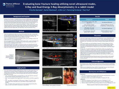
Evaluating Bone Fracture Healing Utilizing Novel Ultrasound Modes, X-Ray and Dual-Energy X-Ray Absorptiometry in a Rabbit Model
Priscilla Machado, Rachel Blackman, Ji-Bin Liu, Flemming Forsberg, and Traci Fox
Background and Purpose
Healing in bone fractures is a complex process involving local inflammation, angiogenesis (i.e., the creation of new blood vessels), and formation of a bone-building callus, all of which leads to returning the bone nearly to its pre-fracture state. This is the process in a bone where the fracture is clean and the fractured ends oppose each other. Five to 10 percent of fractures are described as non-union, which can lead to poor healing and long-term complications. The purpose of this study was to compare fracture healing in a rabbit model using x-ray, the traditional method for evaluating fracture healing, with ultrasound methods including color Doppler imaging (CDI) and power Doppler imaging (PDI), shear wave elastography (SWE), superb microvascular imaging (SMI), and Dual-Energy X-Ray Absorptiometry (DXA).
-

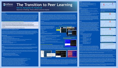
The Transition to Peer Learning
C. G. Roth, H. V. Naringrekar, and A. E. Flanders
Problem Description: Historically, peer review has been compelled by regulatory and legislative mandates, such as the Joint Commission Ongoing Professional Practice Evaluation (OPPE) requirement and the Health Care Quality Improvement Act (HCQIA) enacted by Congress in 1986. [1] However, these external mandates were focused on quality assurance, generally carrying punitive connotations and practically translated into rote compliance without the benefit of learning and improvement. In fact, the lack of quality improvement focus prompted the Institute of Medicine (IOM) to release its 2015 report, “Improving Diagnosis in Health Care,” stating that a “critical type of error in health care—diagnostic error—has received relatively little attention.” [2] The IOM report alarmingly reports that 5% of the US population experience diagnostic error annually, most experience diagnostic error in the course of a lifetime and diagnostic error contributes to 10% patient deaths and 6-17% of adverse events in hospitals. The IOM report framed a number of recommendations that potentially informs peer review and learning activities more broadly (Figure 1).
-

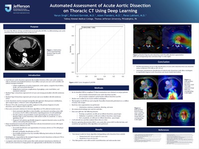
Automated Assessment of Acute Aortic Dissection on Thoracic CT Using Deep Learning
Varun Singh; Richard Gorniak, MD; Adam Flanders, MD; and Paras Lakhani, MD
Purpose
To assess the efficacy of deep convolutional neural networks (DCNNs ) in differentiating acute aortic dissections from non-dissected aortas on thoracic CT.
-

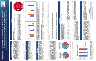
Improving Bedside Procedural Safety through Optimizing Timeout Documentation and a Pre-procedure Checklist
Jennifer Harris, MD; R. Benson Jones, MD; Kristin Lohr, MD; Grant Turner, MD; Drew Kotler, MD; Justine Blum, MD; Megan Margiotta, MD; Matthew Bokhari, MD; Erica Li, MD; Riti Kanesa-thasan, MD; Bracken Babula, MD; and Rebecca Jaffe, MD
Aim
GOAL: Improve the safety of patients undergoing bedside procedures while maintaining the full spectrum of graduated autonomy in procedure training for residents.
SMART Aim: Increase the rate of timeouts documented for bedside procedures from 29% to 50% by June 2018.
-

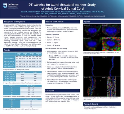
DTI Metrics for Multi-site/Multi-scanner Study of Adult Cervical Spinal Cord
Devon M. Middleton, PhD; Joshua Fisher, BS; Adam E. Flanders, MD; Feroze Mohamed, PhD; John H. Wu, MD; Laura Krisa, PhD; Mark Elliot, PhD; Scott H. Faro, MD; and Jessica Evans, BS
Background and Objective
A major variable in DTI spinal cord studies is the diversity in MRI scanner vendor and field strength. While there are several techniques proven to acquire DTI data, there are no standardized accepted methods for acquisition and processing. As more medical systems are utilizing this technology to evaluate the spinal cord, it is important to study the reproducibility of the DTI metrics among various scanner platforms, coil configurations and software implementations to determine the variance in obtaining normative spinal cord DTI data. This preliminary data for a multi-site DTI study examines the effects of these different MR vendors and field strengths on the DTI values of the adult cervical spinal cord.
-
, CNMT, NMTCB(CT); Cheryl Rickley, CNMT; Charles Intenzo, MD; and Sung Kim, MD")
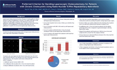
Preferred Criterion for Deciding Laparoscopic Cholecystectomy for Patients with Chronic Cholecystitis Using Radio-Nuclide Tc99m Hepatobiliary Mebrofenin
Duy Tran, R.T(N), CNMT, NMTCB(CT); Cheryl Rickley, CNMT; Charles Intenzo, MD; and Sung Kim, MD
Objective
To determine what gastroenterologist prefer for laparoscopic cholecystectomy when using Tc99m hepatobiliary scintigraphy.
-

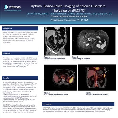
Optimal Radionuclide Imaging of Splenic Disorders: The Value of SPECT/CT
Cheryl Rickley, CNMT; Michele Barasch, CNMT; Charles Intenzo, MD; and Sung Kim, MD
Objective:
Dedicated radionuclide imaging of the spleen in useful in conditions such as accessory spleen and splenic trauma. Nuclear Medicine plays a key role in the diagnostic molecular SPECT/CT imaging of splenic disorders.
Poster presented at Society of Nuclear Medicine Molecular Imagining in Denver Colorado, United States.
-

The Added Value of SPECT/CT in Endocrinology: A Pictorial Essay
Cheryl Rickley, CNMT; Michele Barasch, CNMT; Charles Intenzo, MD; and Sung Kim, MD
Objective:
Dedicated radionuclide imaging of the spleen in useful in conditions such as accessory spleen and splenic trauma. Nuclear Medicine plays a key role in the diagnostic molecular SPECT/CT imaging of splenic disorders.
Poster presented at SNMMI Annual Conference in Denver Colorado, United States.
-

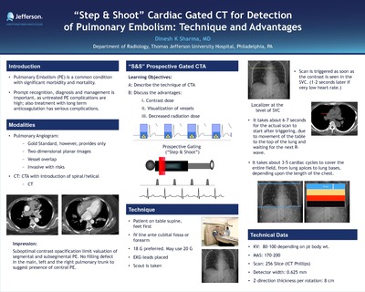
“Step & Shoot” Cardiac Gated CT for Detection of Pulmonary Embolism: Technique and Advantages
Dinesh K. Sharma, MD
Introduction
Pulmonary Embolism (PE) is a common condition with significant morbidity and mortality. Prompt recognition, diagnosis and management is important, as untreated PE complications are high; also treatment with long term anticoagulation has serious complications.
Poster presented at The 11th Congress of Asian Society of Cardiovascular Imaging in Kyoto Japan.
-

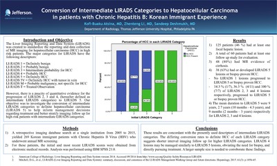
Conversion of Intermediate LIRADS Categories to Hepatocellular Carcinoma in patients with Chronic Hepatitis B: Korean Immigrant Experience
Kofi-Buaku Atsina, MD; Zhenteng Li, MD; and Sandeep Deshmukh, MD
Introduction and Objective
The Liver Imaging Reporting and Data System (LIRADS) was created to standardize the reporting and data collection of MR imaging for hepatocellular carcinoma (HCC) in high risk patients. The major categories for LIRADS have the following descriptors:
LI-RADS 1 = Definitely benign
LI-RADS 2 = Probably benign
LI-RADS 3 = Intermediate probability for HCC
LI-RADS 4 = Probably HCC
LI-RADS 5 = Definitely HCC
LI-RADS 5V = Definitely HCC with tumor in vein
LI-RADS M = Probable malignancy, not specific for HCC
LI-RADS T = Treated Observation
However, there is a paucity of quantitative evidence for the progression of LIRADS 2, 3 and 4, (hereafter defined as intermediate LIRADS categories) to LIRADS 5. Our objective was to investigate the conversion of intermediate LIRADS categories to definite hepatocellular carcinoma (LIRADS 5) to help inform management decisions regarding treatment and better stratify imaging follow up for high-risk patients with intermediate LIRADS categories.
Poster presented at: SCBT-MR in Salt Lake City, Utah.
-

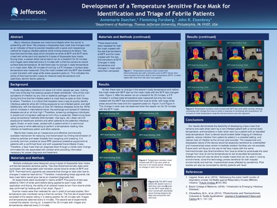
Development of a Temperature Sensitive Face Mask for Identification and Triage of Febrile Patients
Annemarie Daecher, Flemming Forsberg, and John R. Eisenbrey
Abstract:
Many infectious diseases are most transmittable when the carrier is presenting with fever. We propose a disposable face mask that changes color as an indicator of fever to provide hospitals with a quick and inexpensive method for triaging infected patients while limiting exposure to others. Two blue thermochromatic dyes which transition to white at 89°F and 92°F were mixed with white paint and applied to 5 types of disposable face masks. During trials, a wearer either sat at rest or ran on a treadmill for 20 minutes and images were obtained every 5 minutes with a thermal camera to record surface body temperature. At rest there was no change in body temperature or in mask color. After 20 minutes of running, a 5°F increase in surface body temperature was measured. Only the mask created with the 89°F dye showed a color transition with large white areas apparent post-run. This indicates the ability of thermochromatic masks to measure body temperature and potentially identify febrile patients.
-

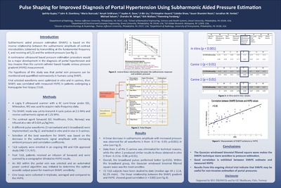
Pulse Shaping for Improved Diagnosis of Portal Hypertension Using Subharmonic Aided Pressure Estimation
Ipshita Gupta, John R. Eisenbrey, Maria Stanczak, Anush Sridharan, Jaydev K. Dave, Ji-Bin Liu, Christopher Hazard, Colette Shaw, Susan Shamini-Noori, Jonathan M. Fenkel, Michael Soluen, Chandra Sehgal, Kirk Wallace, and Flemming Forsberg
Subharmonic aided pressure estimation (SHAPE) is based on the inverse relationship between the subharmonic amplitude of contrast microbubbles (obtained by transmitting at the fundamental frequency fo and receiving at fo/2) and the ambient pressure (see fig.1).
A noninvasive ultrasound based pressure estimation procedure would be a major development in the diagnosis of portal hypertension and less invasive than the current catheter-based hepatic venous pressure gradient (HVPG) measurement.
The hypothesis of this study was that portal vein pressures can be monitored and quantified noninvasively in humans using SHAPE.
First selected waveforms were optimized in vitro and in canines, then SHAPE was correlated with measured HVPG in patients undergoing a transjugular liver biopsy (TJLB).
-
 Subharmonic Aided Pressure Estimation for Monitoring Neoadjuvant Chemotherapy Response of Breast Cancer by Kibo Nam, Maria Stanczak, Anush Sridharan, Adam C. Berger, Tiffany Avery, John R. Eisenbrey, and Flemming Forsberg")
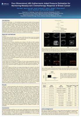
Four-Dimensional (4D) Subharmonic Aided Pressure Estimation for Monitoring Neoadjuvant Chemotherapy Response of Breast Cancer
Kibo Nam, Maria Stanczak, Anush Sridharan, Adam C. Berger, Tiffany Avery, John R. Eisenbrey, and Flemming Forsberg
Introduction
Neoadjuvant chemotherapy is a well-established treatment option for patients with non-metastatic breast cancer. The patient response is correlated with survival. However, the optimal method for monitoring neoadjuvant therapy response has not been established. One factor that may affect the response of neoadjuvant therapy is the interstitial fluid pressure (IFP). Increased IFP prevents an effective delivery of therapeutic agents and reduces the efficacy of the therapy. Recently, subharmonic-aided pressure estimation (SHAPE) using contrast-enhanced ultrasound (CEUS) has been developed and its potential was demonstrated in animals as a non-invasive technique for IFP measurements. The SHAPE method estimates IFP based on the inverse relationship between the subharmonic signal magnitude from CEUS and IFP. The purpose of this study was to determine if 4D SHAPE can predict the response of breast cancer to neoadjuvant chemotherapy.
Poster presented at International Ultrasonics Symposium in Tours France.
Printing is not supported at the primary Gallery Thumbnail page. Please first navigate to a specific Image before printing.
