

Files
Download Full Text (2.5 MB)
Description
Background and Purpose
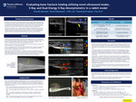
Healing in bone fractures is a complex process involving local inflammation, angiogenesis (i.e., the creation of new blood vessels), and formation of a bone-building callus, all of which leads to returning the bone nearly to its pre-fracture state. This is the process in a bone where the fracture is clean and the fractured ends oppose each other. Five to 10 percent of fractures are described as non-union, which can lead to poor healing and long-term complications. The purpose of this study was to compare fracture healing in a rabbit model using x-ray, the traditional method for evaluating fracture healing, with ultrasound methods including color Doppler imaging (CDI) and power Doppler imaging (PDI), shear wave elastography (SWE), superb microvascular imaging (SMI), and Dual-Energy X-Ray Absorptiometry (DXA).
Publication Date
4-16-2024
Keywords
bone fracture, ultrasound, SMI, SWE
Disciplines
Medicine and Health Sciences | Radiology
Recommended Citation
Machado, Priscilla; Blackman, Rachel; Liu, Ji-Bin; Forsberg, Flemming; and Fox, Traci, "Evaluating Bone Fracture Healing Utilizing Novel Ultrasound Modes, X-Ray and Dual-Energy X-Ray Absorptiometry in a Rabbit Model" (2024). Department of Radiology Posters. 12.
https://jdc.jefferson.edu/radiologyposters/12


Comments
Presented at the 2024 JCHP Collaborative Capstone & Research Exchange.